

KFU biologists offer new insights into markers of amyotrophic lateral sclerosis
Scientists at Kazan Federal University’s Institute of Fundamental Medicine and Biology have found that changes associated with amyotrophic lateral sclerosis (ALS) begin before symptoms appear. Their findings were published in the International Journal of Molecular Sciences.
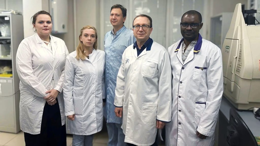
The research team was led by Ekaterina Martynova, a leading research associate at the Gene and Cell Technologies Laboratory. The work was conducted in collaboration with Kazan State Medical University with support from a Russian Science Foundation grant.
The study involved several stages. The researchers used transgenic SOD1-G93A mice, a well-established model of familial ALS carrying a mutation in the superoxide dismutase 1 gene. The animals were divided into groups representing different disease stages, ranging from 30 days of age at the presymptomatic stage to 120 days at the terminal stage.
“We used advanced techniques, including light microscopy of semithin sciatic nerve sections. We also used transmission electron microscopy (TEM) to examine the ultrastructure of the sciatic nerve and the gray matter in the ventral horns of the spinal cord, along with immunofluorescence profiling of the spinal cord using the markers GFAP for astrocytes, IBA1 for microglia, NG2 for NG2 glial cells, and Olig2 for oligodendrocytes. Another key technique was multiplex analysis of serum cytokines and chemokines,” Martynova said.
The team found that peripheral nerve degeneration begins at the presymptomatic stage, with disintegration of the axonal cytoskeleton and thickening of the myelin sheath.
“At later stages, nerve tissue is replaced by connective tissue, indicating a profibrotic shift in Schwann cells. In the spinal cord, we identified two phases of microglial activation: an early neuroprotective phase followed by a neurotoxic phase at the terminal stage, triggered by signals from astrocytes. The area of degenerated tissue increased twelvefold between the presymptomatic and symptomatic stages. We also detected stage-specific changes in serum chemokines: MCP-3 increased at the symptomatic stage, while CTACK, eotaxin, and TECK increased at the terminal stage. These findings suggest that MCP-3 could be a potential early biomarker and therapeutic target in ALS,” she said.
The study supported the concept of distal axonopathy, in which the earliest changes occur in the peripheral portions of neurons rather than in their cell bodies.
“Even at the presymptomatic stage, axonal transport is impaired, the axonal cytoskeleton becomes fragmented, and mitochondrial function declines, resembling Wallerian degeneration. We found that, following denervation, Schwann cells separate into two populations: some become phagocytic, while others undergo a profibrotic shift, replacing nerve tissue with collagen. This pattern has not previously been described in ALS in this form,” Martynova explained.
She also outlined the team’s plans for future research, “Our future work will focus on the intricate mechanisms underlying neurodegeneration. We plan to assess therapeutic approaches using the approved drug edaravone, as well as gene- and cell-based approaches involving human umbilical cord blood mononuclear cells genetically modified with various adenoviral vectors expressing growth and neurotrophic factors. Targeted metabolomic profiling is another promising avenue.”